SKIN PUNCH BIOPSY FOR SMALL FIBER NEUROPATHY
Epidermal Nerve Fiber Density (ENFD) testing is a highly sensitive, skin biopsy, used to identify Small Fiber Neuropathy (SFN). SFN is a disorder of the peripheral nerves that affect small somatic fibers, autonomic fibers, or both, resulting in sensory changes and or autonomic dysfunction when both types of fibers are involved. ENFD testing is the most reliable tool and considered the “Gold Standard for Testing” when diagnosing SFN. ENFD testing provides a definitive diagnosis, which assists the provider in treating and monitoring the prescribed regimen.
ENFD Testing was developed by Johns Hopkins and University of Minnesota. There are 100’s of articles speaking to its reproducibility and acceptance in medical diagnostics. The biopsy is minimally invasive and performed using a 3 mm punch tool. The patient’s skin is anaesthetized with 2% lidocaine with epinephrine, which alleviates the pain during the biopsy. The procedure takes the provider approximately 90 seconds to perform, with the entire procedure taking about 10 -15 minutes. Samples are placed in Zamboni tubes and shipped in pre-paid packaging. The biopsy sites are covered with a bandage and do not require any special treatment.
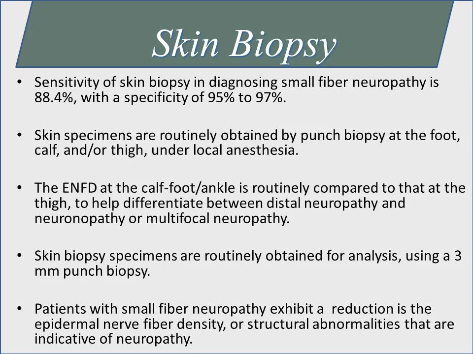
The diagnostic efficiency of skin biopsy is approximately 88%. For diagnosing SFN, it is more sensitive than quantitative sensory testing and more sensitive and less invasive than sural nerve biopsy. ENFD provides a definitive diagnosis for SFN. Aggressive cause-specific treatment, lifestyle modifications, and pain control are key elements of managing SFN. Patients presenting with pain, numbness, and paresthesia, are appropriate for ENFD testing. Additionally, patients with diabetes mellitus, prediabetes, metabolic syndrome, fibromyalgia, autoimmune diseases, alcohol abuse, and idiopathic neuropathy are candidates, based on their presenting symptomatology. We offer an assessment survey to assist in the screening process to identify appropriate patients for the skin punch biopsy.
Confirming the diagnosis of SFN will allow the provider to evaluate possible underlying etiologies efficiently, and aggressively manage the symptoms. Etiology-specific treatment is crucial in preventing SFN and or slowing its progression. Glucose control, weight control, well-balanced diets, regular exercise, physical therapy, vitamin supplements, prescription drugs, avoiding exposure to toxins and limiting or avoiding alcohol are components of the treatment regimen for SFN.
COMMON OBJECTIONS:
“I would never do it because it would not change my treatment options.”
“This is only beneficial to Neurology offices.”
“I consider it more academic and a waste of money.”
People want medical care that involves both diagnosis and treatment. People want to know what's wrong with them especially in pain management as many have been told "it's all in your head.” It verifies to that person that they have a real disease even if the treatment options are the same. However, treatment options are not the same depending on the cause. The first-line and best treatment would be to identify and treat the underlying cause whether it be pre-diabetes/DM, hypothyroidism, hyperlipidemia/metabolic syndrome, or an infectious disease such as HIV/Hep C which are all independent causes of small fiber neuropathy. Treatment of the underlying cause may not eliminate all of the pain but will improve the person's pain symptoms and slow the progression of the disease and save a significant amount of money in unnecessary imaging studies and trial and error medication choices that do not address the underlying cause. From a medical-legal standpoint, if someone has undiagnosed disease such as the ones stated above which could be easily diagnosed with post-procedure blood work and the patient’s condition worsens that physician could be implicated if a malpractice claim was initiated.
Simple example: A patient comes into the clinic with extremely high blood pressure and a headache. You wouldn't just give some aspirin and send them home. You would treat the underlying cause which is the high blood pressure. It's the same for small fiber neuropathy. You need to treat the cause not just the symptoms.
“I do not see the value in the biopsies because I would rather do an EMG because the reimbursement is higher and then if it is negative then I would just treat the symptoms.”
Skin biopsy testing for small fiber neuropathy does not preclude EMG/NCV testing. EMG/NCV tests the large myelinated A-alpha fibers but does not test the small myelinated A-delta and unmyelinated C-fibers which transmit pain and temperature sensations. One test does not preclude the other and performing both would be recommended.
TYPES OF NEUROPATHY THAT CAN BE DETECTED:
Diabetic Neuropathy
Peripheral Neuropathy is one of the most common microvascular complications of diabetes.
Roughly 50% of diabetics suffer from PN.
50% of these neuropathies are considered at least moderate in severity.
Histological studies suggest primarily small C fibers are affected by diabetes and glucose intolerance.
Orstavik, K., et al., Abnormal function of C-fibers in patients with diabetic neuropathy. J Neurosci, 2006. 26(44): p. 11287-94.
Pittenger, G.L., et al., Intraepidermal nerve fibers are indicators of small-fiber neuropathy in both diabetic and nondiabetic patients. Diabetes Care, 2004. 27(8): p. 1974-9.
Polydefkis, M., et al., The time course of epidermal nerve fibre regeneration: studies in normal controls and in people with diabetes, with and without neuropathy. Brain, 2004. 127(Pt 7): p. 1606-15.
Smith, A.G., et al., Epidermal nerve innervation in impaired glucose tolerance and diabetes-associated neuropathy. Neurology, 2001. 57(9): p. 1701-4.
Diabetic Neuropathy morbidities amount to more than $10 Billion.
Gordois, A., et al., The health care costs of diabetic peripheral neuropathy in the US. Diabetes Care, 2003. 26(6): p. 1790-5.
Peripheral Neuropathy
Most are “mixed fiber.”
Small, medium, and large fibers can be equally affected.
Electrophysiologically these are detected as axonal sensorimotor neuropathies.
Neuropathies may start as small fiber and evolve into mixed fiber.
This can be seen in diabetes where patients have symptoms but no objective evidence of neuropathy for years.
A small percentage remain confined only to small fibers.
Medication-Induced Neuropathy
Taxanes (Paclitaxel and Docetaxel) are used to treat solid cancerous tumors in the ovary, breast, lung, head and neck.
Promote microtubule stabilization prohibiting mitotic division. This can lead to spontaneous demyelination and inhibits regenerative capacities of the neurons.
Years of opioid or narcotic pain killer use can lead to neuropathic problems. As well as missing nerve degenerative problems do to pain being masked for too long.
Should be noted that there is a marked predisposition to drug-induced neuropathy in diabetic patients.
Kus, T., et al., Taxane-induced peripheral sensory neuropathy in cancer patients is associated with duration of diabetes mellitus: a single-center retrospective study. Support Care Cancer, 2016. 24(3): p. 1175-9.
Alcoholic Neuropathy
Occurs in 2/3 of Chronic Alcoholics. Combinational Multi-Fiber Neuropathy.
Large Fiber Damage likely attributed to Thiamine deficiency.
Small Fiber Damage likely related to toxic intermediate metabolites, Aldehydes (Acetaldehyde) and Ketones.
Oxidative stress can also lead to DNA fragmentation and neuron apoptosis.
SFN in Fibromyalgia
27 patients with fibromyalgia and 30 matched normal controls were studied. They found that:
41% of skin biopsies from subjects with fibromyalgia vs 3% of biopsies from control subjects were diagnostic for SFN
8 subjects had dysimmune markers, 2 had hepatitis C serologies, and 1 family had apparent genetic causality.
These findings suggest that some patients with chronic pain labeled as fibromyalgia have unrecognized SFN.
TREATMENT OF NEUROPATHY
Diabetic Control
18 months of lowering HgA1c scores patients showed decreased pain, and increased nerve fiber count on ENFD.
Exercise Alone
Most important element of lifestyle improvement; patients showed increase in ENFD count.
Alpha Lipoic Acid
Potent Antioxidant (coenzyme in Krebs Cycle)
Reduces micro and macro vascular complications
600 mg IV – reduces neuropathy symptoms immediately
600 mg PO qd, b.i.d., t.i.d. – reduces symptoms (has GI side effects)
Trental (Pentoxifylline) 400 mg q8h
Changes shape and flexibility of red blood cells so the red blood cells can reach the nerves in the capillaries.
Relaxes smooth muscle for vasodilation
Helps wound healing
Prevents cellulitis
May cause GI upset
Topamax (Topiramate) – Has shown nerves to grow back after 6 weeks on ENFD counts
Nitric Oxide Supplements
L-Citrulline 4 grams t.i.d.
L-Arginine 1 gram t.i.d.
Thiamine
SKIN BIOPSY PROCEDURE
In-Office Procedure that can be performed by any clinician. Specimens cannot be processed in regular pathology lab so once specimens are taken they must be sent to a specialized lab for processing. Sensitivity at Gentox is reported to be roughly 90%. Values lower than normal indicate diagnosis of small fiber neuropathy. Normal fiber count yet large swelling in nerves indicate early disease.
Two to four 3mm sites are chosen. Process should only take 10-15 minutes.
Distal Leg
Distal Thigh
Proximal Thigh
Podiatrists can use:
10 cm Proximal to Fibula, Sural Nerve
Extensor Digitorum Brevis Muscle Belly, Superficial Fibular Nerve
Clean Biopsy Area with alcohol pad or alternate antiseptic cleansing pad.
Inject Lidocaine in an apex proximal pattern around biopsy site. DO NOT INJECT DIRECTLY OVER THE BIOPSY SITE!!!!
Once the patient’s site has been fully anesthetized, wait 5-10 minutes before proceeding.
The 3mm punch is then inserted onto the biopsy site and gently rotated, letting the blade do the cutting.
Keep instrument perpendicular to skin surface.
Insert to level of sub-cutis.
If necessary, gently use forceps to remove the tissue.
Place into labeled Zamboni tubes. Ensure the epithelium is NOT DAMAGED during this process.
Clean biopsy site once more. Place white gauze or Band-Aid over the biopsy site. No stitches necessary. Heals up as a small scar with no special treatment. Most cases the site is not even visually detectable within a few months post exam.
Each biopsy must be labeled with Name, DOB and Site of Biopsy.
Place into the Biohazard Bag:
Biopsy Specimen
Completed Work Order
Place into the Shipment bag:
Ice-Pack inserted into the sleeve
Biohazard Bag
Call local FedEx for pickup
Biopsy immune-stained with a Panaxonal Marker (PGP9.5)
Small Nerve Fibers in the epidermis counted under a microscope.
Intraepithelial Nerve Fiber Densities calculated & compared to normal values.


