


0
items
$0
CONE BEAM COMPUTED TOMOGRAPHY (CB CT) SCANNER
The PreXion3D Excelsior CBCT Scanner is the evolution of our original flagship product designed for dentists who demand the highest diagnostic clarity and detail. Uses include but are not limited to: implant placement, bone quality studies, pathology, periodontics and endodontics. It is compatible with all DICOM compliant third-party implant surgical guide and CAD/DICOM merging software programs. All PreXion3D CBCT scanners include The PreXion3D Viewer software at no additional charge. It contains easy modules for evaluating dedicated 2D Panoramic, 2D Cephalometric, and 3D images. PreXion provides the clinician with the most accurate assessment of the bone and surrounding anatomy while making 1:1 exact measurements. This assures optimal implant placement without the superimposition of tissue or projection distortion compared to conventional panoramic systems. PreXion3D reconstructs to DICOM 3.0 format and is compatible with major third-party surgical planning software systems.
Better diagnose patients with more detail and clarity
Present cases more confidently, increase acceptance
Create the wow factor with patients
We are committed to your success by focusing on low dose and advanced imaging technologies for accurate diagnosis and treatment. PreXion’s legacy has always been founded on providing superior imaging quality with powerful and easy to use software. With over 15 years of experience in 3D DICOM image processing and software visualization, PreXion has distinct advantages over other CBCT scanners and is committed to “Make IT Visible” under every clinical circumstance.
Improving image quality with exclusive innovative technologies. PreXion has improved the reliability and quality of all CBCT core technologies including the X-Ray Tube and the Flat Panel Detector (FPD) for both CT and Cephalometric. All done in-house, PreXion is supplying superior quality products from our state of art factory in Japan with excellence in quality assurance, production, and manufacturing flexibility. Our newly developed FPD, X-Ray Tube, and software – coupled with the world’s smallest focal spot of 0.3mm – deliver industry leading image clarity with 6 times larger volume rendering for every clinical need. PreXion 3D Excelsior enables CBCT imaging with 30% lower patient radiation dose than current PreXion CBCT levels without any degradation in image quality.
Increase your diagnostic and treatment accuracy with optimal FOV size PreXion 3D Excelsior has 5 distinct FOV sizes ranging from 50x50mm, 100x50mm, 100x81mm, 150x81mm and 150x130mm, perfect to select for any clinical indication. Variation of imaging mode and the possibility to select the volume size according to your diagnosis allow you to obtain the highest image quality, all while by limiting radiation (in accordance with ALARA principles).
5x5 images work well for a specific area diagnosis with minimal X-ray exposure for the patient. It is especially effective for endodontic diagnosis by checking root canal conditions. 10x5 images generate a wide range to view oral conditions in the maxillary and mandibular areas. It is especially effective for clinical procedures related to the mandibular nerve, mental foramen, or maxillary sinus. 10x8 images generate comprehensive diagnosis and treatment planning. It is effective for complex implant surgery and diagnosis of the TMJ. 15x8 images generate the most optimal information for oral diagnosis covering both maxillary and mandibular structures in a single scan. It is effective for most oral surgery cases including placement of multiple implants. 15x13 images generate the most optimal information of larger implant cases, airway and TMJ analysis, orthodontics and dental sleeping solutions. (Vertical Stitched)
Versatility of selection. PreXion 3D Excelsior provides several scan modes on the CT scan, Panoramic scan, Cephalometric exposure.
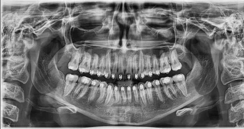
THE ADVANCED IMAGING QUALITY WITH NEW STANDARD OF PANORAMIC IMAGE. A clear and sharp panoramic image generates better diagnostics. Advanced details, especially in the anterior and dental roots can be easily viewed with PreXion 3D Excelsior. These consistently high quality panoramic images are the new standard of panoramic imaging solutions.


Capturing every specialty. PreXion 3D Excelsior is designed to serve as an expandable platform to offer Cephalometric options for all your orthodontic needs. PreXion 3D Excelsior provides optimal images with an exclusively designed and developed FPD for cephalometric diagnosis. Our one-shot Cephalometric sensor delivers short exposure time with an incredibly low dose for the patient. The large 7” touchscreen with an easy-to-use interface enables simple usage and set up. The user-friendly controls allow for quick, effortless, and intuitive workflow for all staff members for any clinical procedure. Being able to directly and immediately position the patient has never been easier. The ability for the clinician to perfectly align the gantry for any patient regardless of height or size is quickly accomplished with a visual guide on the 7” touchscreen located directly beside the patient. Intuitive and efficient usability defined.


ORTHODONTICS
Proper orthodontic treatment starts with proper diagnosis. The PreXion Excelsior shows the exact relationship of all anatomy in stunning clarity for your treatment planning and case presentation. With our intuitive software, cephalometric tracing and measurements are fast and easy – further enhancing case planning with precision and accuracy. With the Excelsior’s ultra-low dose mode, you can capture all the information you need with minimal radiation exposure, ensuring peace of mind for you and your patients.
IMPLANTS
The use of CBCT in implant dentistry is arguably the most common of all clinical applications. With the advancements in image quality found in the PreXion Excelsior, the highest levels of predictability and clinical success are at your fingertips. With our powerful implant planning software, you can easily and quickly map the mandibular nerve, assess bone quantity and quality, and determine the proper implant angulation and position. In addition, you now have the capability to export DICOM and STL data for further planning and surgical guide fabrication.
ORAL SURGERY
The ability to see what you previously have not been able to is invaluable to any practitioner who is either performing the surgery, or collaborating with a surgeon on a referral basis. Clearly see third molars, supernumerary teeth, and maxillary cuspids. Properly diagnose cysts, tumors, and lesions. Identify sinus walls, nerve positioning, and other vital structures and locations. The PreXion Excelsior will enhance outcomes and minimize complications.
PERIODONTICS
The PreXion Excelsior has direct impact on all aspects of a progressive periodontal practice. From applications in implant planning and treatment, the ability to evaluate bone loss and defects, diagnose disease and pathology – there is not an area that the clarity of the Excelsior image cannot bring into focus and make visible.
ENDODONTICS
The ability of the PreXion Excelsior to capture small areas with high resolution is one of the most powerful capabilities we can offer to endodontic diagnosis and treatment. The ability to identify an endodontic lesion and abscess and the capacity to see canals not visible in 2D are but a few of the capabilities unlocked with the Excelsior.
TMJ
The PreXion Excelsior is able to aid in the assessment of the TMJ by showing condyle and fossa morphology in extremely clear detail. Diagnose and evaluate potential TMD issues including identifying erosions, osteophytes, and fractures. Accurately measure the volume and the surface of the condyle. With the PreXion Excelsior’s dedicated TMJ Scan mode, your patients are exposed to a fraction of the radiation.
AIRWAY ANALYSIS
With the PreXion Excelsior you can discover restricted airways, and see the pathway for optimal treatment for patients suffering from sleep disorders, sleep apnea, and sinus complications. Unlock the ability with our software to easily trace the airway, see restriction points, and calculate volume.
ENT / OTOLARYNGOLOGY
In a position paper by The American Academy of Otolaryngology-Head and Neck Surgery (approved 4/13/2010 revised 9/28/2013) they stated that CBCT is an appropriate imaging modality of the paranasal sinuses, skull base, and temporal bones due to the fact that CBCT provides greater spatial resolution with significantly lower radiation levels than conventional CT (medical CT, MRI, or standard radiography). The ability to provide point-of-care CBCT for diagnosis to otolaryngology patients has far-reaching and life altering benefits - immediate review of 3D images, a reduction in patient anxiety, immediate diagnosis, higher patient compliance, and reduced medical costs. Every medical and dental specialty deserves the best. Treatment starts with diagnosis – now you can clearly see both health and disease of the sinonasal area, the temporal bone, maxillofacial trauma and abnormalities, and the inner ear with a fraction of the radiation with incredible image clarity. Make It Visible.